Home
/ Lower Body Skeletal Anatomy - Lower Extremity Anatomy Bones Muscles Nerves Vessels Kenhub : The heart and lungs are located within the thoracic cavity, and the vertebral column provides structure and protection for the spinal cord.
Lower Body Skeletal Anatomy - Lower Extremity Anatomy Bones Muscles Nerves Vessels Kenhub : The heart and lungs are located within the thoracic cavity, and the vertebral column provides structure and protection for the spinal cord.
Lower Body Skeletal Anatomy - Lower Extremity Anatomy Bones Muscles Nerves Vessels Kenhub : The heart and lungs are located within the thoracic cavity, and the vertebral column provides structure and protection for the spinal cord.. Since the spine is encircled by musculature, the abdomen, spinal muscles, and hips are all integral aspect of maintaining a healthy lower spine and therefore lower back. Every skeletal muscle has three main parts: Discussed in this article as part of the axial skeleton is a third subdivision, the visceral, comprising the lower jaw, some elements of the upper jaw, and the branchial arches, including the hyoid bone. The leg is specifically the region between the knee joint and the ankle joint. The lumbar spine makes up the the lower end of the spinal column.
The tibia, or shin bone, spans the lower leg, articulating proximally with the femur and patella at the knee joint, and distally with the tarsal bones, to form the ankle joint. It consists of the bones that make up the arms and legs, as well as the bones that attach them to the. The lower limb contains 30 bones. The brain is surrounded by bones that form part of the skull. The structural framework of the hip region is provided by the pelvis, a structure composed of the pelvic girdle and the coccyx.
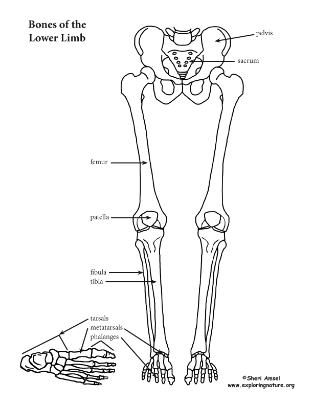
Lower Limb Thigh Leg And Foot from www.exploringnature.org The action refers to the action of each muscle from the standard anatomical position. Tibia and fibula in anatomical position with parts labeled. The skeleton protects vital organs. Discussed in this article as part of the axial skeleton is a third subdivision, the visceral, comprising the lower jaw, some elements of the upper jaw, and the branchial arches, including the hyoid bone. Distal to the ankle is the foot. The lower limb contains 30 bones. The back muscles are skeletal muscles. The brain is surrounded by bones that form part of the skull.
The thigh is that portion of the lower limb located between the hip joint and knee joint.
This curve, called lordosis, helps to: The muscles of the lower back, including the erector spinae and quadratus lumborum muscles, contract to extend and laterally bend the vertebral column. It consists of the skull, vertebral column (including the sacrum and coccyx), and the thoracic cage, formed by the ribs and sternum. The muscles of the human body can be categorized into a number of groups which include muscles relating to the head and neck, muscles of the torso or trunk, muscles of the upper limbs, and muscles of the lower limbs. It consists of 5 lumbar vertebra that are numbered 1 through 5 from top to bottom i.e. The l5 vertebra is connected to the top of. Anatomical drawings 12 photos of the anatomical drawings anatomical drawings 17th century, anatomical drawings definition, anatomical drawings of insects, anatomy drawings tutorial, leonardo da vinci anatomical drawings exhibition, human anatomy, anatomical drawings 17th century, anatomical drawings definition, anatomical drawings. Balance the weight of your head on top of your spine evenly distribute weights from your upper body into the lower extremities The back muscles are skeletal muscles. The brain is surrounded by bones that form part of the skull. Interactions between the skeleton, muscles, and nerves move the body. In turn, the pelvic girdle consists of two hip bones and the sacrum, interconnected at the pubic symphysis and sacroiliac joints. Appendicular skeleton anatomy there are a total of 126 bones in the appendicular skeleton.
Browse 222 lower back skeleton stock photos and images available, or start a new search to explore more stock photos and images. Balance the weight of your head on top of your spine evenly distribute weights from your upper body into the lower extremities The lower spine, the hips and tailbone, and the abdomen. Appendicular skeleton anatomy there are a total of 126 bones in the appendicular skeleton. The back muscles are skeletal muscles.
1 Appendicular Skeletal Anatomy Primate Appendages Consist Of A Download Scientific Diagram from www.researchgate.net Human anatomy diagrams show internal organs, cells, systems, conditions, symptoms and sickness information and/or tips for healthy living. The muscles of the lower back help stabilize, rotate, flex, and extend the spinal column, which is a bony tower of 24 vertebrae that gives the body structure and houses the spinal cord. They support bones, in this case, the vertebrae. The structural framework of the hip region is provided by the pelvis, a structure composed of the pelvic girdle and the coccyx. The lower back is really composed of three areas of the body: Related posts of anatomy of lower body anatomical drawings. The skeletal system also provides attachment points for muscles to allow movements at the joints. It provides structure to the body, and each bone has a distinct purpose.
Skeletal muscular system of the lower body.
The lower limb contains 30 bones. The skeleton of the lower limbs and the vertical spinal column is a unique evolutionary device that allowed a person to raise his head above all other living creatures on our planet. The l5 vertebra is connected to the top of. The anatomy of the lumbar spine is quite complex. It provides structure to the body, and each bone has a distinct purpose. The collection of bones in the human body is called the skeletal system. Here we will attempt to provide a brief overview of lumbar spinal anatomy. It consists of 5 lumbar vertebra that are numbered 1 through 5 from top to bottom i.e. The lower limb skeleton includes the cingulum membri inferioris (the lower limbs bones belt) and skeleton membri inferioris liberi. Skeletal muscular system of lower body (pdf download) skeletal muscular system of lower body (streaming audio) all course materials are streaming audio & digital files and not physical materials. The skeletal system also provides attachment points for muscles to allow movements at the joints. The brain is surrounded by bones that form part of the skull. Discussed in this article as part of the axial skeleton is a third subdivision, the visceral, comprising the lower jaw, some elements of the upper jaw, and the branchial arches, including the hyoid bone.
L1, l2, l3, l4, and l5. By tightening and relaxing, the skeletal muscles create movement. This diagram depicts lower extremity diagram. Related posts of anatomy of lower body anatomical drawings. The lumbar spine makes up the the lower end of the spinal column.
Anatomy Standard Landing Page from www.anatomystandard.com The skeletal system also provides attachment points for muscles to allow movements at the joints. Each bone is a complex living organ that is made up of many cells, protein fibers, and minerals. The muscles of the lower back, including the erector spinae and quadratus lumborum muscles, contract to extend and laterally bend the vertebral column. It consists of the bones that make up the arms and legs, as well as the bones that attach them to the. It consists of the skull, vertebral column (including the sacrum and coccyx), and the thoracic cage, formed by the ribs and sternum. Some, like the rib cage, provide protection for softer body parts, while other bones enable mobility by supporting the muscles. Start studying lower limb skeletal anatomy. The axial skeleton supports the head, neck, back, and chest and thus forms the vertical axis of the body.
This diagram depicts lower extremity diagram.
Appendicular skeleton anatomy there are a total of 126 bones in the appendicular skeleton. This curve, called lordosis, helps to: It consists of skull, vertebral column, and thoracic cage. Human anatomy diagrams show internal organs, cells, systems, conditions, symptoms and sickness information and/or tips for healthy living. The brain is surrounded by bones that form part of the skull. The back muscles are skeletal muscles. The leg is specifically the region between the knee joint and the ankle joint. The action refers to the action of each muscle from the standard anatomical position. Tibia and fibula in anatomical position with parts labeled. Some, like the rib cage, provide protection for softer body parts, while other bones enable mobility by supporting the muscles. The axial skeleton forms the axis of the human body. The skeleton acts as a scaffold by providing support and protection for the soft tissues that make up the rest of the body. Related posts of anatomy of lower body anatomical drawings.

